Author Archives: Tom Bouthillet

Transcutaneous Pacing (TCP) With and Without Capture

EMS is dispatched to a private residence for 70-year-old female who is believed to be unconscious.

On arrival, the patient is found lying in bed unresponsive to painful stimuli. The patient’s skin is pale and clammy. Her shirt is damp. Snoring respirations are noted and a slow carotid pulse is present.

A medical history is obtained from family members and includes heart failure, stroke, and hypertension.

Vital signs are assessed.

  • RR: 8
  • HR: Less than 30
  • NIBP: 78/41
  • SpO2: Not registering
  • Temp: 96.1 F / 36.7 C
  • BGL: 101 mg/dL

The cardiac monitor is attached.

High degree AV block with wide complex escape rhythm.

The adult pads are placed and transcutaneous pacing is initiated.

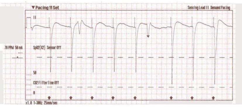
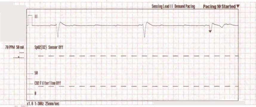
The transcutaneous pacer is set for 70 PPM at 50 mA. Pacing spikes are visible with what appear to be large, corresponding QRS complexes.

The patient’s blood pressure improves slightly to 84/47 (confirmed by auscultation). However, paramedics are still concerned about the patient’s hypotension.

Additional treatments

  • IO access is obtained in right proximal tibia.
  • 0.5 mg of Atropine is administered x 3.
  • 9% normal saline is run wide open with an additional IV line established in the left lower extremity.

The patient begins to move and reaches for the pacing pads. However, she is still non-verbal and does not follow commands.

  • On arrival at the hospital the patient is transitioned to transvenous pacing.
  • She is sent to the cardiac cath lab where a permanent pacemaker is placed.
  • In the ICU the patient remains dangerously hypotensive in spite of dobutamine and levophed drips.

The patient eventually expires from multiple-system organ failure.

Discussion

Transcutaneous pacing (TCP) is a difficult skill that is often performed incorrectly. The problem of false capture (also known as echo distortion) is under-recognized and under-reported in the medical literature.

There are many reasons why medical professionals often fail to achieve true electrical and mechanical capture. Consider this excerpt from the Journal of Emergency Medicine where Douglas Ettin, M.D. and Thomas Cook, M.D. describe the difficulty.[1]

“Shortly after cardiac pacing was initiated, the patient’s systolic blood pressure dropped to 50 mmHg. The EKG monitor continued to demonstrate adequate ventricular capture by the pacer. The patient appeared to have palpable pulses; however, the rhythm contractions of the patient’s body from the pacer shocks made this assessment difficult. With the etiology of the patient’s hypotension unclear, the decision was made to use transthoracic ultrasonography to assess the adequacy of her ventricular contractions.”

“Initially, the ultrasound demonstrated ventricular contractions at a rate of 30-40 beats per minute. These heart contractions did not correspond with the surrounding thoracic muscle contractions generated by the pacer. The current was gradually increased to 110 mA, and the heart began to contract in unison with the pacer shocks. The patient’s blood pressure subsequently increased to 90 mmHg.”

You can see another example where an echo was used to verify capture here.

With false capture, you will generally see a near-vertical upstroke or down-stroke to the “phantom” QRS complex (which is actually electrical artifact created by the current passing between the pacing pads).

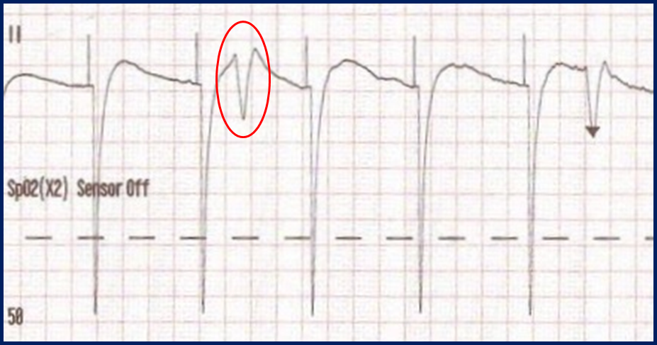
Classic “false capture” with near-vertical down-stroke of the (phantom) QRS complexes, slightly curved return to the isoelectric line, and unimpressive T-waves.

You will also note that the underlying rhythm can be seen in the absolute refractory period of one of the (presumed to be) paced QRS complexes (red circle). That is not scientifically possible!

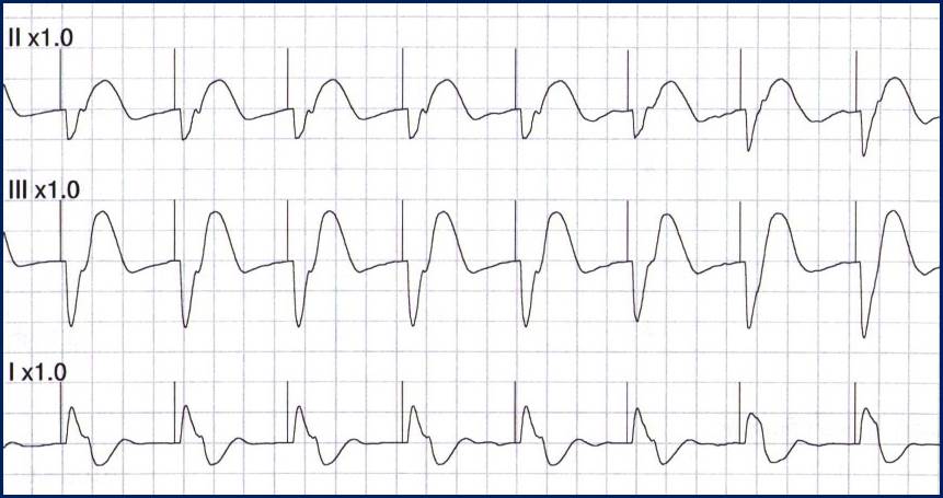
In contrast, true electrical capture will show wide QRS complexes with tall, broad T-waves.

Transcutaneous pacing (TCP) with true electrical capture as evidenced by tall, broad T-waves. Capture was achieved at 110 mA (ems12lead.com).

Tips for success

  • Perform, but do not rely on a pulse check!
  • Use an instrument (SpO2, Doppler, capnography, or echo) to help confirm mechanical capture whenever possible
  • Do not be fooled by skeletal muscle contraction!
  • Know that the patient may become more alert whether capture is achieved or not
  • The most common reasons for “failure to capture” are insufficient milliamperes and poor pad placement!

More examples of transcutaneous pacing (TCP) with capture

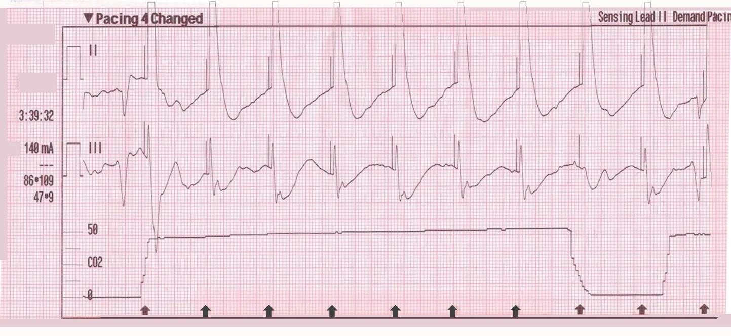
Capture achieved at 140 mA and confirmed with sudden rise in ETCO2 (ems12lead.com).

110 mS

Capture achieved at 110 mA confirmed with SpO2 plethysmograph

Reference

  1. Ettin DCook T. Using ultrasound to determine external pacer capture. The Journal of Emergency Medicine. 1999;17(6):1007-1009.

Further reading

Pacing Artifact May Masquerade As Capture

This case was submitted by Roger Hancock with edits by Tom Bouthillet. Some details have been changed to protect patient confidentiality.

Syncope with Acute Inferior STEMI and 3 Different AV Blocks

This is a case from the “old days” when prehospital 12-lead ECGs were still a bit of a novelty. Many of the details have been lost to time but patient’s heart rhythms will be the focus of this review.

The patient was a young woman in her late 40s who presented with syncope while playing tennis. Syncope during exercise is troubling and suggests a possible cardiac cause, which is potentially life threatening.

EMS arrived on scene and obtained a detailed history. The woman admitted to some chest discomfort. Vital signs were assessed and the cardiac monitor was attached.

Recorded ECG #1

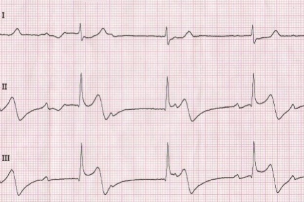
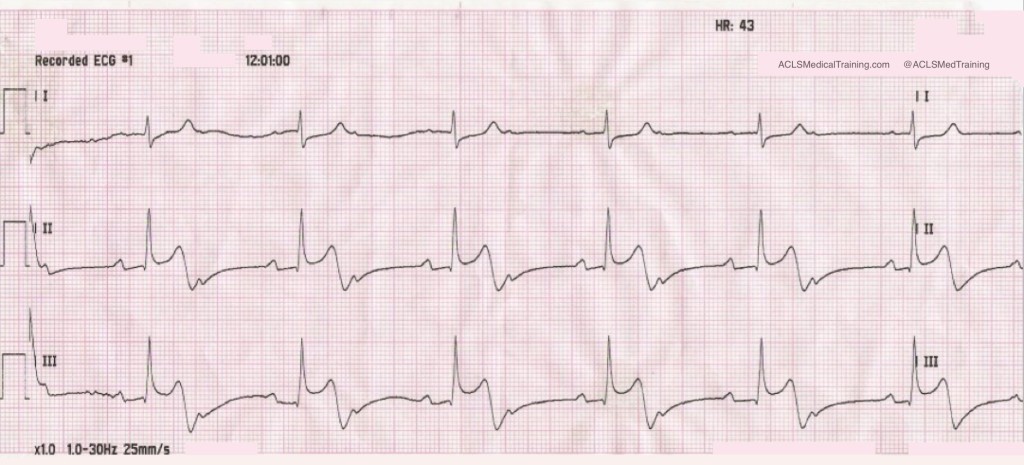
The initial ECG showed second degree AV block with 2:1 conduction.

This is often called second degree AV block type 2 with 2:1 conduction but second degree AV block with 2:1 conduction is untypeable.

There appears to be an acute injury pattern even though the rhythm strip is recorded in ‘monitor’ mode with the low frequency / high pass filter set to 1.0 Hz.

A few minutes later a rhythm change was noted on the monitor.

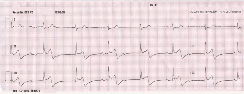
Recorded ECG #2

Now the rhythm is third degree AV block with junctional escape rhythm. The atrial rate is about 60 and the ventricular rate is 41.

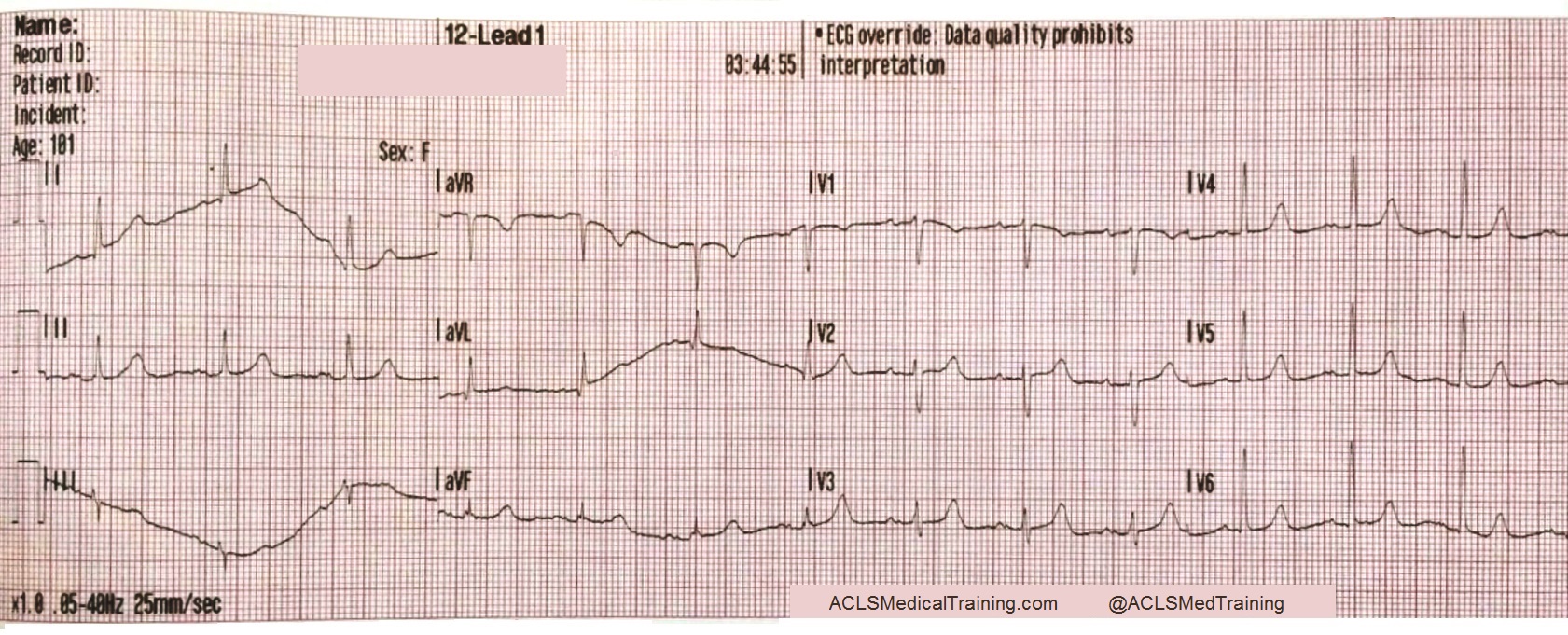
(The initial 12-lead ECG showed poor data quality but paramedics understood that the patient was suffering acute STEMI.)

Aspirin was given and an IV was started. Nitroglycerin was also given and the patient became hypotensive.

Remember, this case is over 10 years old. At that time there was a lot less emphasis on things like right ventricular infarction and the cardiac cath lab was not activated based on the prehospital 12-lead ECG.

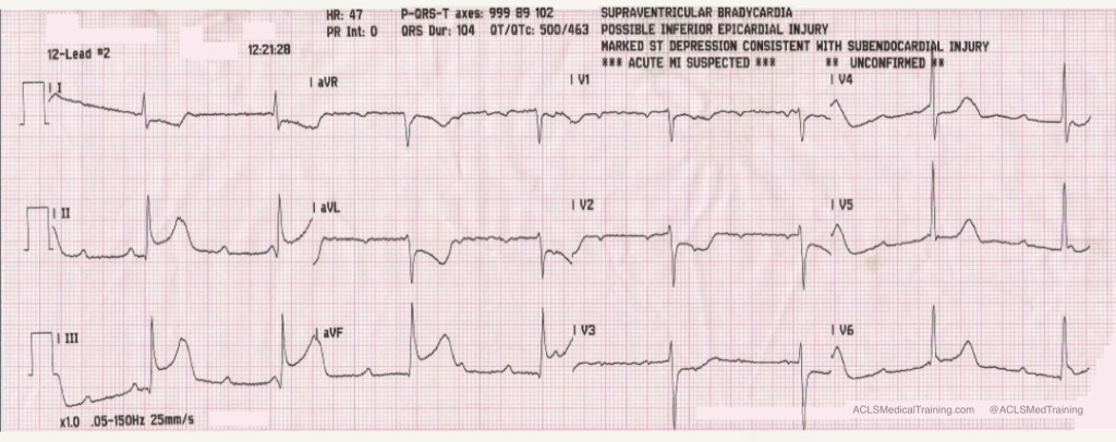
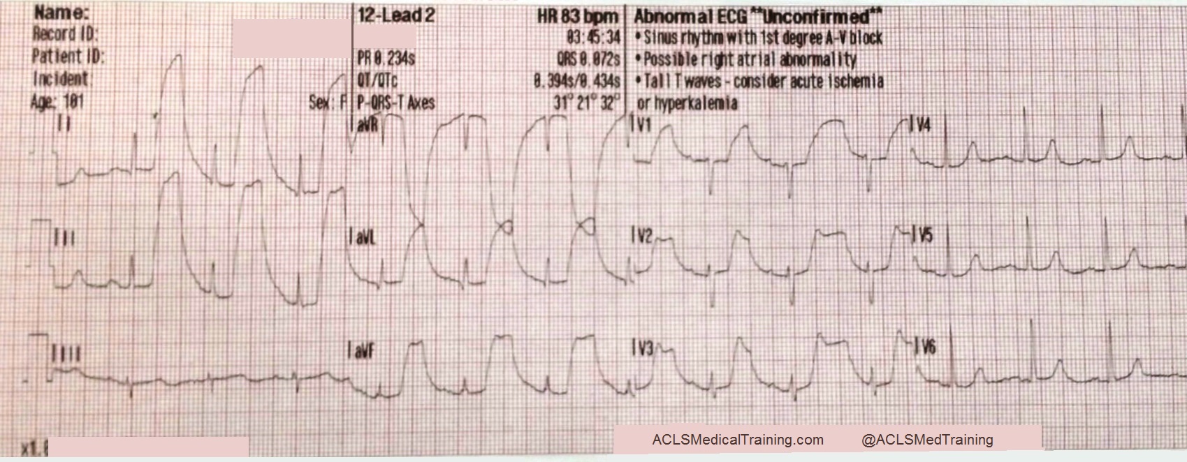
0.5 mg atropine was given rapid IVP and another 12-lead ECG was obtained.

12-Lead #2

The atrial rate has doubled to about 125. The ventricular rate has increased modestly to about 47. The ECG is diagnostic for acute inferior-posterior STEMI.

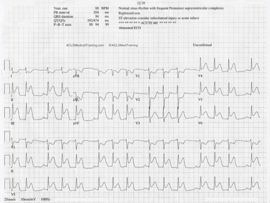
On arrival in the Emergency Department the staff obtained their own 12-lead ECG.

Second degree AV block type 1

Now the heart rhythm is second degree AV block type 1 (Wenckebach).

How can you tell? In the first place we see clustering of QRS complexes (huge tip-off) and we already know that AV conduction is precarious. The initial cardiac cycle of each cluster shows a constant PR-interval.

Progressive Prolongation

When we take a closer look at the rhythm strip we see progressive prolongation of the PR-interval until a P-wave is “dropped” proving that the heart rhythm is second degree AV block type 1 (Wenckebach).

The ECG shows worsening of the ST-segment elevation. The patient was sent to the cardiac cath lab. As far as I know she made a full recovery.

Discussion

Heart blocks in the setting of acute STEMI can result either from ischemia of the AV node or increased parasympathetic tone, which is a manifestation of the Bezold-Jarisch reflex.

Consider these excerpts from Braunwald’s Heart Disease (Fifth Edition). It’s an old book but it contains some interesting information.

“The AV conduction system has a dual blood supply, the AV branch of the RCA and the septal perforating branch from the LAD. Therefore, complete heart block can occur in patients with either anterior or inferior infarction. Complete heart block develops in 5 to 15% of all patients with AMI; the incidence may be even higher in patients with RV infarction. As with other forms of AV block, the prognosis depends on the anatomical location of the block in the conduction system and the size of the infarction.”

“Complete heart block in inferior infarction usually results from an intranodal or supranodal lesion and develops gradually, often progressing from first degree or type I second degree block. The escape rhythm is usually stable without asystole and often junctional, with a rate exceeding 40 beats/min and a narrow QRS complex in 70% of cases and a slower rate and wide QRS in the others […] The mortality may approach 15% unless RV infarction is present, in which case the mortality associated with complete AV block may be more than doubled.”

“[P]atients with inferior MI and AV block have larger infarcts and more depressed right ventricular and left ventricular function than do inferior infarcts with no AV block. As already noted, junctional escape rhythms with narrow QRS complexes occur commonly in this setting…”

“Only when complete heart block develops in less than 6 hours after the onset of symptoms is atropine likely to abolish the AV block or cause acceleration of the escape rhythm. In such cases the AV block is likely to be transient and related to increases in vagal tone rather than the more persistent block seen later in the course of MI, which generally requires cardiac pacing.”

Guide to Understanding ECG Artifact

Artifact on the electrocardiogram can result from a variety of internal and external causes from Parkinsonian muscle tremors to dry electrode gel.

Most of the time it will be obvious that you are dealing with artifact and troubleshooting the problem will be straight forward. However, there are occasions when artifact mimics ECG abnormalities that can cause problems for patient care.

Once when I was a cardiac monitoring technician the alarm sounded and it appeared as though ventricular tachycardia was on the monitor. When we rushed to the patient’s room it turned out she was brushing her teeth!

With a trained eye you can often learn to spot the underlying rhythm “marching” through this type of artifact. Other times it’s not that easy (PDF).

Here are some types of artifact you may encounter along with some tips to help you achieve excellent data quality on your ECG tracings.

Loose lead artifact

You will frequently encounter loose lead artifact when dealing with patients who are diaphoretic because the electrodes simply will not stick to the patient’s body. You may also see this type of artifact when placing the electrode over hair.

To troubleshoot this problem make sure you prep the skin carefully!

Consider tincture of benzoin. It works great for diaphoretic patients. However, tincture of benzoin is flammable! You don’t want to use it for defibrillation pads.

In this example loose lead artifact can be seen in leads I and II.

Loose lead artifact

What electrode do leads I and II have in common?

  • Lead I is a dipole with the negative electrode on the right arm and the positive electrode on the left arm.
  • Lead II is a dipole with the negative lead on the right arm and the positive electrode on the left leg.

Lead I and lead II share the right arm electrode! That is the electrode that is causing this problem.

Wandering baseline artifact

Wandering baseline artifact presents as a slow, undulating baseline on the electrocardiogram.  It can be caused by patient movement, including breathing.

Wandering baseline artifact

I have also noticed that stopping or accelerating the ambulance can cause wandering baseline. Some references suggest that wandering baseline can be caused by loose or dry electrodes.

Some paramedics ask patients to hold their breath while they capture a 12-lead ECG. I don’t do this because it can alter the patient’s heart rate.

There are times when your patient is acutely short of breath and it’s simply impossible to capture a 12-lead ECG with excellent data quality.

Muscle tremor artifact

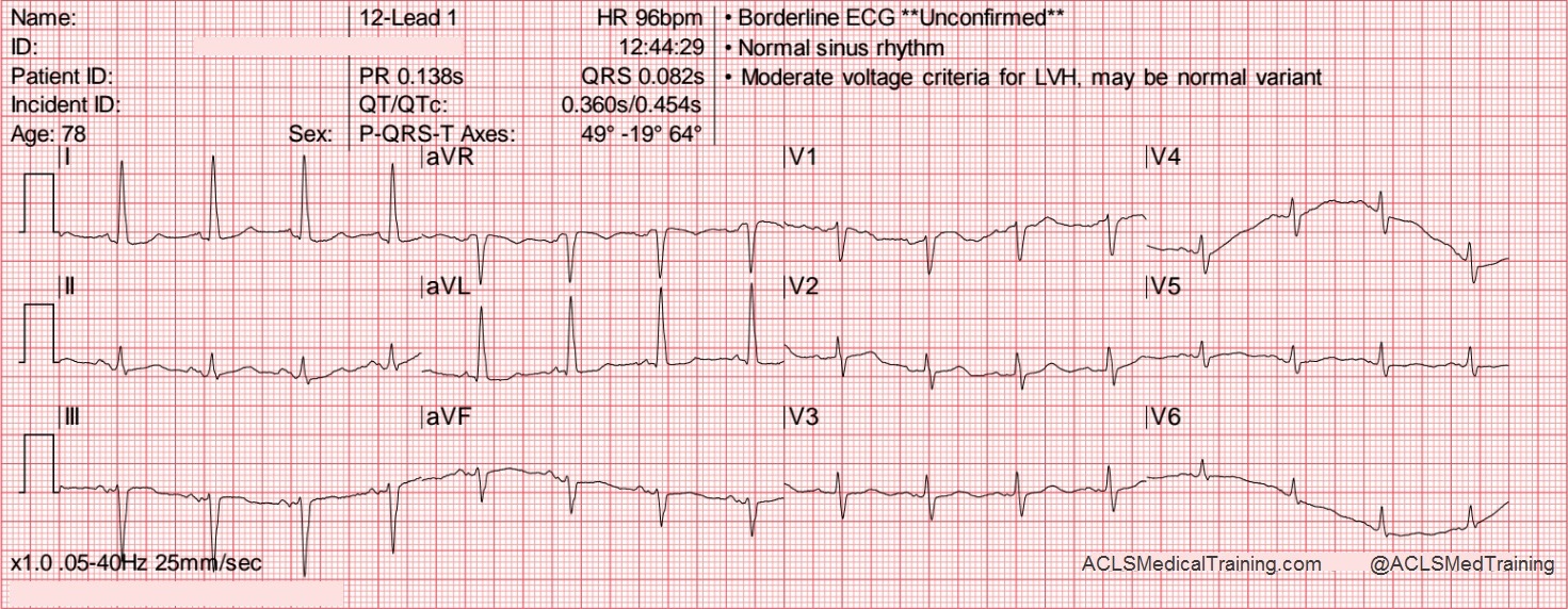
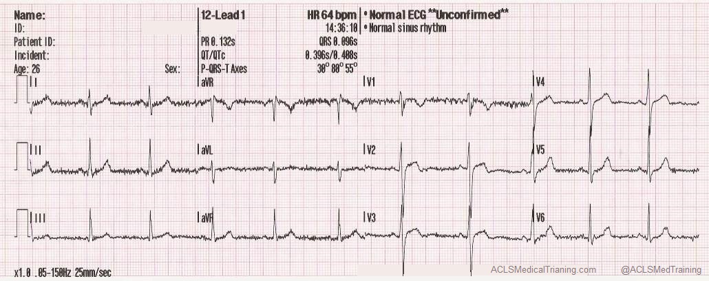
Muscle tremor (or tension) artifact is a type of motion artifact. Usually it’s happening because your patient is cold and shivering. However, it can also happen when patients prop themselves up by their arms.

The example below was obtained from a young, healthy firefighter during routine training. It was cold in the fire station and he was shivering.

12-Lead 1

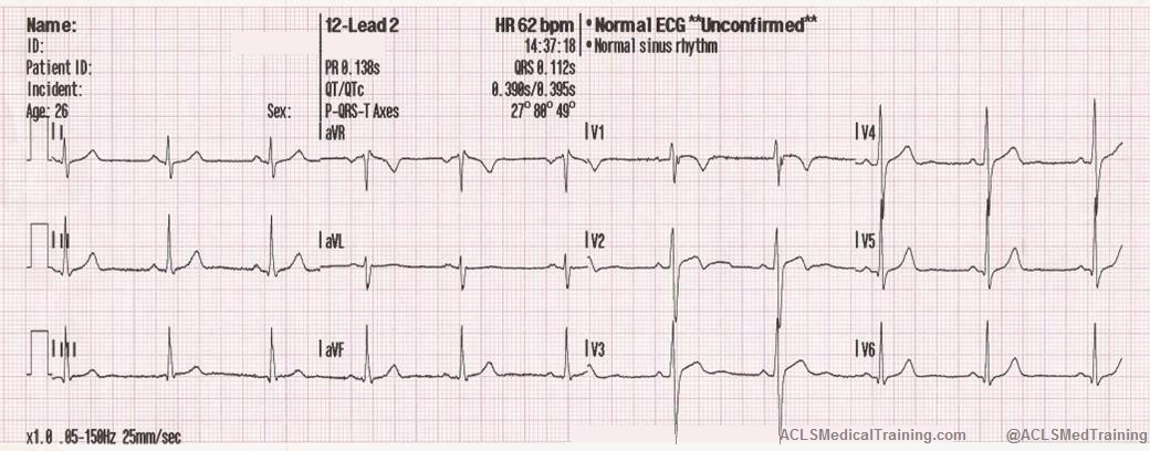
The next example was taken after a large towel was placed over the firefighter. It made quite a difference didn’t it?

12-Lead 2

Electromagnetic interference (EMI)

Electromagnetic interference (EMI) artifact usually results from electrical power lines, electrical equipment, and mobile telephones. In the United States this is sometimes referred to as 60 cycle interference (or 60 Hz pickup).

Electromagnetic interference (EMI)

Creative Commons: ECGGuru.com

To help minimize 60 cycle interference you can set the diagnostic mode of your 12-lead ECG monitor to 0.05 – 40 Hz. As long as the low frequency / high pass filter (the lower number) is set to 0.05 Hz you should get accurate ST-segments.

My department has been doing this for so long that I don’t have any good examples of 60 cycle interference!

CPR compression artifact

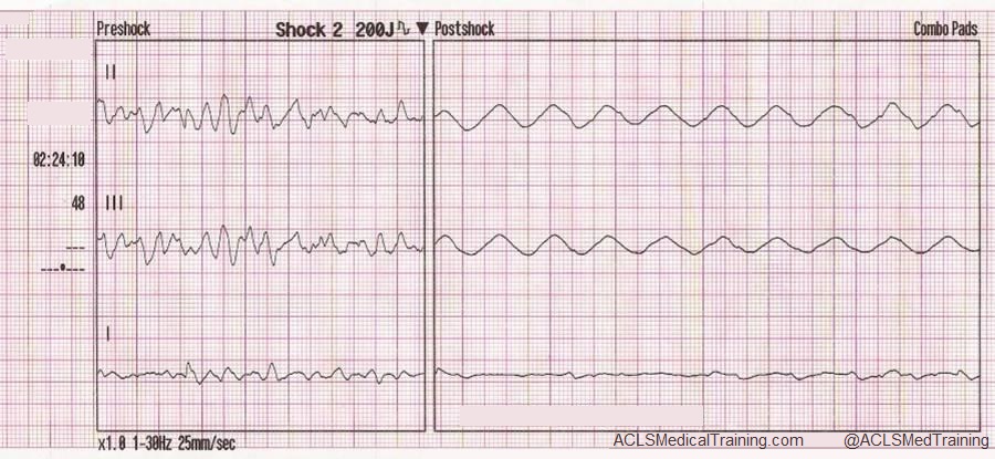
This ECG was automatically recorded during a cardiac arrest.

Preshock and Postshock

The wavy line after the shock is CPR artifact. Using the small block method (1500/13=115) we can determine that the compression rate was about 115/min. which is perfect!

There may be times when CPR artifact makes it difficult to determine the underlying rhythm. However, if you’re performing CPR at a 30:2 compression to ventilation ratio you can see the underlying rhythm during ventilations!

Neuromodulation artifact

Here’s a type of artifact we’re starting to see more frequently as implantable neurostimulators become more prevalent.

These devices are used to treat a variety of symptoms including tremors, seizures, chronic pain, nausea and vomiting related to gastroparesis, problems with bladder or bowel control, visual impairment, and hypertension.

EKG artifact

If you see artifact that looks like this you should ask your patient if he or she has any implantable medical devices. Some devices can be temporarily turned off with a magnet but you should consult with the prescribing physician.

Echo distortion artifact

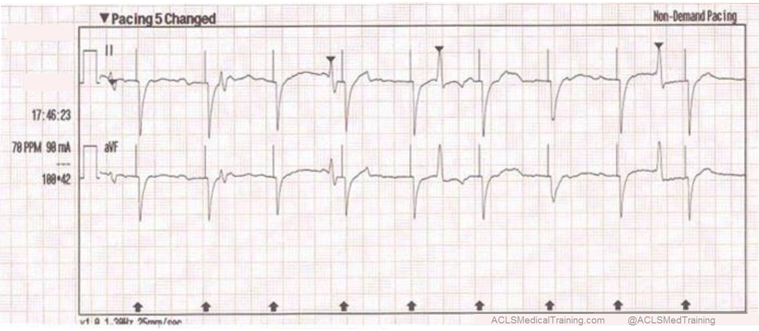
This type of artifact is associated with transcutaneous pacing (TCP). Echo distortion causes a pseudo-QRS complex after the pacing spike which is sometimes referred to as “false capture.”

Pacing 5 Changed

The pacing spike is a graphical representation that electrical current is about to pass between the pacing pads. It is followed by a short “blanking period” of about 40 ms (one small block) where the monitor essentially “closes its eyes”. If it did not, the signal would go right off the ECG paper!

After the blanking period the monitor “opens its eyes” to see the QRS complex that is created by the pacing stimulus. However, sometimes the monitor catches the pacing current as it returns to baseline causing a pseudo-QRS complex on the ECG.

You can read more about the problem of false capture here.

Arterial pulse tapping artifact

This unusual artifact causes large, bizarre T-waves on the ECG. The phenomenon was first reported in 2005 by Özhan et al. as a “bizzare electrocardiogram” thought to be associated with abnormal left ventricular motion.

Subsequent work by Aslanger solved the issue in favor of arterial pulse tapping (which explains why the artifact occurs synchronously with the cardiac cycle on the ECG.)

Consider these two ECGs which were recorded from the same patient less than 1 minute apart. The first ECG shows simple motion artifact in leads I, III, and aVL.

Simple motion artifact

Courtesy of Frank Intessimoni (@njmedic3228)

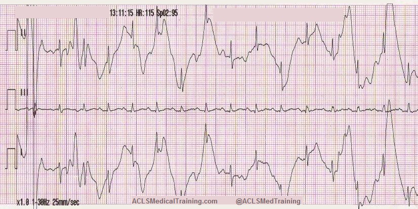
The second ECG shows large, bizarre T-waves that were concerning to the paramedics on the call.

Bizarre T-waves

Courtesy of Frank Intessimoni (@njmedic3228)

You will note that the artifact is most pronounced in leads I, II, and aVR. Lead III appears perfectly normal. This suggests that the right arm electrode was placed over the radial artery.

But if that’s true why is there also artifact in other leads?

Aslanger explains:

“[O]ne may expect that the leads not connected to the electrode affected by the source of disturbance would be free of distortion; but this is not the case. When one of the limb electrodes is affected by a source of disturbance, it distorts not only the corresponding derivation but also [the others] which are all calculated by mathematical equations…”

“…precordial leads [are also affected] because the Wilson central terminal, which constitutes the negative pole of the unipolar leads, is produced by connecting 3 limb electrodes via a simple, resistive network to give an average potential across the body.”

References
Aslanger E, Yalin K. Electromechanical association: a subtle electrocardiogram artifact. Journal of Electrocardiology. 2012;45(1):15-17. doi:10.1016/j.jelectrocard.2010.12.162.
Aslanger E, Bjerregaard P. Mystery of “bizarre electrocardiogram” solved. Journal of Electrocardiology. 2011;44(6):810-811. doi:10.1016/j.jelectrocard.2011.04.001.

Treating Supraventricular Tachycardia with Adenosine

EMS is called to a local medical clinic for a 53 year old female complaining of weakness and palpitations.

Symptoms started earlier in the day at tennis camp. The patient experienced one other episode about 2 years prior that proved to be self-limiting.

She takes no medications and has no known drug allergies.

The patient appears anxious but is oriented to person, place, time, and event.

Vital signs are assessed:

  • HR: 200
  • NIBP: 134/102
  • RR: 18
  • Temp: 98.4 F
  • SpO2: 95% on RA
  • BGL: 88

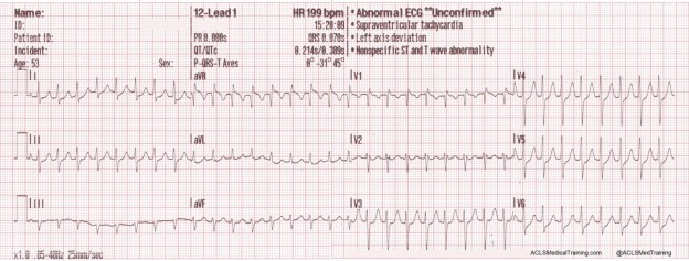
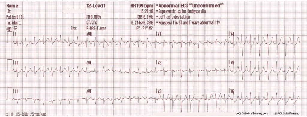
A 12-lead ECG is obtained by paramedics.

A 12-lead ECG is obtained by paramedics

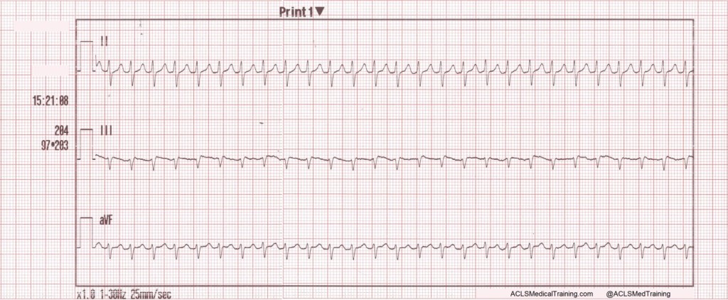
A rhythm strip is also recorded.

A rhythm strip is also recorded

Paramedics note a regular narrow complex tachycardia at a rate of about 200/min.

Could this be sinus tachycardia?

It is doubtful. There are no visible P-waves. In addition, the maximum theoretical sinus rate is 220 minus age (plus or minus 10%). For this patient that works out to somewhere between 167 and 184.

Vagal maneuvers are attempted but are unsuccessful.

As a side note, the REVERT Trial which was published this year introduced a postural modification (leg elevation and supine positioning) to the standard Valsalva maneuver for the treatment of SVT which returned 40% of patients to sinus rhythm compared with 17% for the standard Valsalva maneuver.

You can watch a video of the technique here: https://www.youtube.com/watch?v=8DIRiOA_OsA

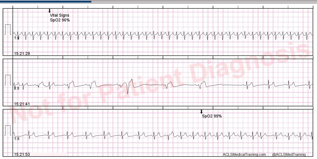
In this case, an IV is started and 12 mg of adenosine is given rapid IV push followed by a 20 ml syringe bolus of 0.9% normal saline.

Vital Signs

The rhythm is successfully converted and another 12-lead ECG is obtained.

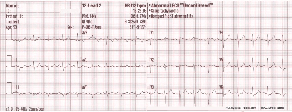
12-Lead ECG

It should be noted that modest nonspecific ST/T wave abnormalities are not uncommon immediately following the conversion of SVT to sinus rhythm. The main determinant of myocardial oxygen demand is heart rate!

Take-away points

The maximum theoretical sinus rate is 220 minus age (plus or minus 10%).

Adenosine should be used for regular tachycardias only! It can be dangerous in the setting of atrial fibrillation and Wolff-Parkinson-White Syndrome.

Record a 12-lead ECG whenever possible prior to treating a narrow complex tachycardia with adenosine. It can be helpful later on when the patient is referred to a cardiologist or electrophysiologist.

Consider a postural modification (leg elevation and supine positioning) to the Valsalva maneuver to improve the conversion rate.

Consider applying defibrillation pads prior to the administration of adenosine.

The drugs Dipyridamole (Persantine) and Carbamazepine (Tegretol) can potentiate adenosine.

Dispatcher

2015 AHA ECC Guidelines – Part 2

This is the second post of a multi-part series reviewing the 2015 AHA ECC Guidelines.

Reference Part 5: Adult Basic Life Support and Cardiopulmonary Resuscitation Quality

Epidemiology of sudden cardiac arrest

The guidelines make this sobering observation: 70% of out-of-hospital cardiac arrests occur in the home and 50% are unwitnessed. Only 10.8% of patients who receive resuscitative efforts by EMS survive to hospital discharge.

My department’s survival rate for unwitnessed cardiac arrest is 0% and I suspect that’s the case for the majority of EMS systems.

Basic life support is still the foundation of resuscitation

The chain-of-survival for out-of hospital cardiac arrest remains unchanged from 2010 with a few updates.

There is a greater emphasis on dispatcher recognition of sudden cardiac arrest with early CPR instructions (sometimes referred to as telephone CPR or T-CPR).

The guidelines reiterate the importance of high quality CPR.

  • Ensuring adequate rate (100-120)
  • Ensuring adequate depth (2 to 2.4” or 5 to 6 cm)
  • Allowing full chest recoil (avoid leaning)
  • Minimizing interruptions to chest compressions
  • Avoiding excessive ventilation

This update recognizes the value of a highly choreographed approach that allows simultaneous approach to chest compressions, ventilations, airway management, rhythm detection, and shocking “by an integrated team of highly trained rescuers in appropriate settings.”

There is an acknowledgement that survival rates for witnessed VF can be as high as 50% in top performing EMS systems but vary from 5% to 50% — a 10 fold difference!

Clearly we would not accept this much variation in operative mortality.

Dispatcher recognition of cardiac arrest and CPR instructions

This may be one of the most important statements in the new guidelines:

“When dispatchers ask bystanders to determine if breathing is present, bystanders often misinterpret agonal gasps or abnormal breathing as normal breathing. This erroneous information can result in failure by dispatchers to identify potential cardiac arrest and failure to instruct bystanders to initiate CPR immediately. An important consideration is that brief, generalized seizures may be the first manifestation of cardiac arrest.”

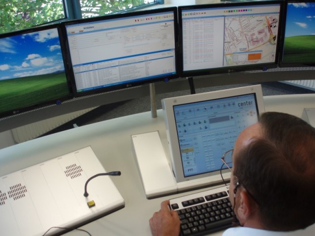
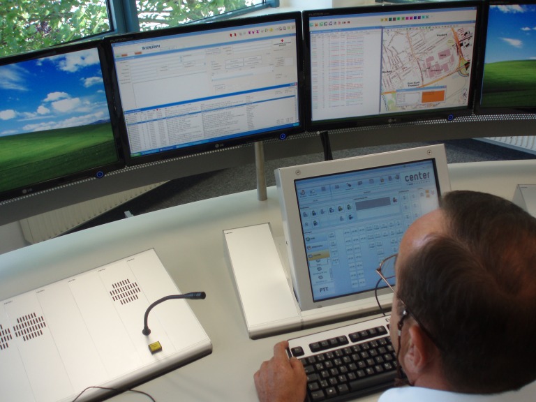
Dispatcher

Image credit: Wikipedia

Quality Improvement in the Dispatch Office

Performing QA/QI on the call intake process for sudden cardiac arrest is extremely important and it’s much harder than you may realize. It is doubtful that the QA/QI tools that come with Emergency Medical Dispatcher (EMD) software packages are adequate.

The CARES registry has an optional T-CPR module that measures various benchmarks for dispatcher performance including recognition of the need for CPR, how long it takes for the dispatcher to begin CPR instructions, and how long it takes for the bystander to begin compressions.

This represents a huge culture shift for dispatch and there is likely to be push back, especially if this type of monitoring occurs without appropriate training, or is implemented in a way that is punitive. However, it’s an often-neglected area of system performance.

As reported by the CARES Registry:

“One recent T-CPR initiative implemented guideline-based protocols, training, and feedback to staff at three regional dispatch centers. This “bundle of care” doubled the number of cases where bystanders started dispatch-directed compressions and reduced the time those compressions started by 80 seconds.”

That’s significant! This is simply not an area we can afford to ignore if we want to be good at resuscitation.

Do we want to be good at resuscitation?

If so, then these issues demand our time and attention. Once you’ve optimized on-scene performance (and hopefully realized some significant gains) you will plateau or perhaps even regress if you don’t find new opportunities to improve the chain-of-survival.

Updated recommendation for dispatchers

“It is recommended that emergency dispatchers determine if a patient is unresponsive with abnormal breathing after acquiring the requisite information to determine the location of the event. If the patient is unresponsive with abnormal or absent breathing, it is reasonable for the emergency dispatcher to assume that the patient is in cardiac arrest. Dispatchers should be educated to identify unresponsiveness with abnormal breathing and agonal gasps across a range of clinical presentations and descriptions.”

Will we pay attention to this recommendation?

I must admit that I find it a bit troubling that so many of my colleagues are saying “there’s nothing new in the 2015 guidelines.” This emphasis on the importance of the call intake process is critically important in my estimation and deserves to be taken seriously.

I hope I’m wrong but it almost reminds me of the 2000 AHA ECC Guidelines when it was first suggested that paramedics may not have enough experience to be proficient at tracheal intubation, and that alternate airways like the LMA might be more appropriate.

We simply did not like the message and many of us still don’t. We’re still arguing about it 15 years later although even the most stubborn of us have, at a minimum, admitted that tracheal intubation should not significantly interrupt chest compressions.

Anecdotally, having reviewed between 50 and 100 cardiac arrest calls using CODE-STAT for CPR analytics, it seems to me that most paramedics will not interrupt chest compressions for more than 10 or 15 seconds for the first attempt. However, if they miss the first attempt, all bets are off, and it is not uncommon for the second attempt to interrupt compressions for 15 to 30 seconds.

Because I know this tends to happen I have asked our paramedics to concentrate on expertly performed BLS for the first 5 cycles (10 minutes) of the cardiac arrest. A 30 second delay in chest compressions, while obviously not desirable, is more palatable at the 10 or 15-minute mark than during the “sweet spot” of the code.

I hope that many years from now (long after I am gone) others in our profession won’t look back and realize that we missed a huge opportunity to strengthen the chain-of-survival in our communities.

Again, I don’t suggest this will be easy. In fact, I’m certain it will be difficult. But with this recommendation it’s a good time to try.

Update: Click here to listen to Robert Lawrence interview Ben Bobrow, M.D. about dispatcher recognition of cardiac arrest and CPR instructions. Here’s the most important part:

“We have to look at this intervention as something we can quantify. So it’s not a binary thing. It’s not a yes or no…it’s not like a 9-1-1 system does this or they don’t, because almost every 9-1-1 system if you ask…answers that they do provide this life-saving service. But when you actually ask them how they train, what protocols they use, if they have a set protocol, how they measure it, what their performance standards are, it’s all over the board. So I think one of the main concepts, besides turning the public into an army of first responders, is that we need to measure, and have performance standards, for giving pre-arrival CPR instructions.”

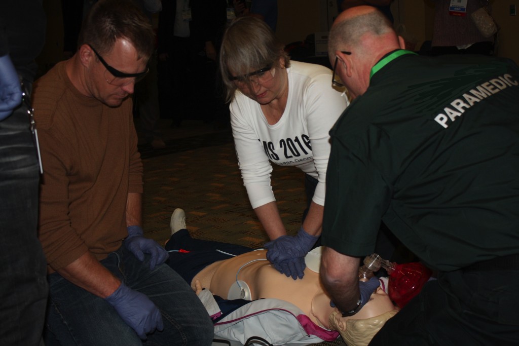
High Performance CPR training at EMS Today 2015

High Performance CPR training at EMS Today 2015


Consideration of the likely cause of the cardiac arrest

I personally found this update to be interesting:

“[I]t is realistic for healthcare providers to tailor the sequence of rescue actions to the most likely cause of arrest. For example, if a lone healthcare provider sees an adolescent suddenly collapse, the provider may assume that the victim has had a sudden arrhythmic arrest and call for help, get a nearby AED, return to the victim to use the AED, and then provide CPR.”

In my mind it is linked to the next issue.

Delayed positive pressure ventilation

Consider these recommendations:

“For witnessed OHCA with a shockable rhythm, it may be reasonable for EMS systems with priority-based, multitiered response to delay positive-pressure ventilation by using a strategy of up to 3 cycles of 200 continuous compressions with passive oxygen insufflation and airway adjuncts.”

And:

“We do not recommend the routine use of passive ventilation techniques during conventional CPR for adults. However, in EMS systems that use bundles of care involving continuous chest compressions, the use of passive ventilation techniques may be considered as part of that bundle.”

Setting aside the qualifiers (we could debate what it means to provide “priority-based, multitiered response” or to use “bundles of care”), the guidelines now acknowledge two important facts.

  • Professional rescuers are capable of discerning between run-of-the-mill sudden cardiac arrest and asphyxial arrest which could potentially change how we approach the situation.
  • Positive pressure ventilation is probably unnecessary during the first 6 minutes for witnessed VF/VT arrest.

This to me is a big change in the guidelines. Even though many EMS systems (including my own) have been using a form of Pit Crew CPR which is a hybrid between cardiocerebral resuscitation and 30:2, it’s nice to see the guidelines officially recognize the practice.

I have a suspicion that switching to continuous chest compressions with “passive oxygen insufflation” for the first 3 cycles (6 minutes or so) has the potential to save a lot of lives, especially in those EMS systems that have been waiting for the AHA to endorse the practice.

Rhythm checks and defibrillation

Rhythm checks and defibrillation

The guidelines reinforce that a defibrillator should be used as soon as possible and that chest compressions should be performed while the defibrillator is “being retrieved and applied.”

The evidence shows no survival benefit for a prescribed interval of 1.5 to 3 minutes of chest compressions prior to the first shock (when compared to a control group where chest compressions are performed while the defibrillator is being prepared).

However, I know from conducting time trials in my own department that it takes a good minute to turn on the defibrillator, extend the cables, attach the pads, apply the pads to the patient, charge the capacitor, and deliver the first shock.

So unless you’re by yourself there’s no reason the patient should not receive 100 chest compressions while all of this is happening.